


Fluorophores and Optical Filters for Fluorescence Microscopy
This is Section 9.3 of the Imaging Resource Guide.
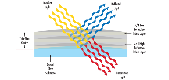
Fluorescence microscopy is a microscopy technique that uses fluorescence, which is induced using fluorophores, as opposed to absorption, scatter, or reflection. A fluorophore (or fluorochrome) is a fluorescent dye used to mark proteins, tissues, and cells with a label for examination by fluorescence microscopy. A fluorophore works by absorbing energy of specific wavelengths, commonly referred to as the excitation range, and re-emitting that energy in another specific wavelength region, referred to as the emission range.
Fluorescence microscopy systems like an epifluorescent microscope can be simple, whereas confocal or multiphoton systems can be very complex. However, all fluorescence microscopes share the same basic
concept: excitation energy illuminates a sample which releases a weak but quantifiable emission energy. The excitation and emission wavelengths do not share the same center wavelength. This allows specialized filters to increase the overall contrast and signal.

Figure 1: A typical fluorescence microscope setup.
The most basic concept and schematic is seen in Figure 1. A filter arrangement is constructed of three filter types: an excitation filter, a dichroic filter, and an emission filter.
Filter #1: Excitation Filter
The excitation filter is placed within the illumination path of a fluorescence microscope and filters out all wavelengths of the light source except for the fluorophore excitation range. The filter minimum transmission dictates the brightness and brilliance of images. A minimum of 40% transmission for any excitation filter is recommended such that the transmission is ideally >85%. The bandwidth of the excitation filter should be entirely within the fluorophore excitation range such that the center wavelength (CWL) of the filter is as close as possible to the peak excitation wavelength of the fluorophore. The excitation filter optical density (OD) dictates the background image darkness; OD is a measure of how well a filter blocks the wavelengths outside of transmission range or bandwidth. A minimum OD of 3.0 is recommended but an OD of 6.0 or greater is ideal.
Filter #2: Dichroic Filter or Beamsplitter
The dichroic filter is placed between the excitation filter and emission filter at a 45° angle and reflects the excitation signal towards the fluorophore while transmitting the emission signal toward the detector. Ideal dichroic filters and beamsplitters have sharp transitions between maximum reflection and maximum transmission, with a >95% reflection for the bandwidth of the excitation filter and a transmission of >90% for the bandwidth of the emission filter. Select the filter with the intersection wavelength (λ) of the fluorophore in mind, to minimize stray-light and a maximize the fluorescent image signal-to-noise ratio.
Filter #3: Emission Filter
The emission filter is placed within the imaging path of the fluorescence microscope and filters out the fluorophore excitation range while transmitting the emission range. The same recommendations for excitation filters hold true for emission filters: minimum transmission, bandwidth, OD, and CWL. An emission filter with the ideal CWL, minimum transmission, and OD combination provides the brightest possible images, with the deepest possible blocking, and ensures the detection of the faintest emission signals.

Figure 2: A generalized fluorophore spectral curve.
Figure 2 shows a typical excitation, emission profile. The absorption and emission profiles share common wavelengths which is one reason why high-quality filters with high transmission, narrow bandwidths, high ODs, and sharp cut-on and cut-off bands are needed. Using low quality filters can ultimately damage the sample, specimen, or expensive sensors. Contact us to help you select the appropriate filters your application.
How Does EO Match Filter Pairs?
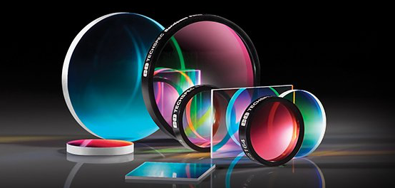
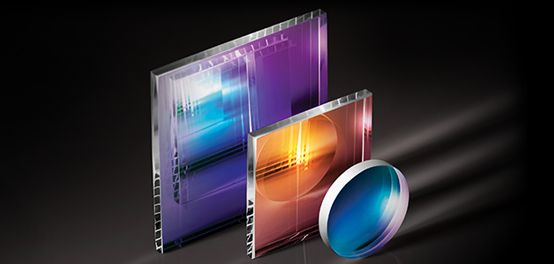
Edmund Optics offers dozens of in-stock and ready to ship optical filters that fit within the excitation and emission regions of each specified fluorophore. We make it easy to quickly search filters by fluorophore. Simply browse the recommended filters that match the peak excitation or emission wavelength, with maximum transmission at that wavelength. For fluorescence microscopy applications using multiple fluorophores, laser sources, alternate dichroic filters or beamsplitters, or applications more complex than typical fluorescence microscope setups, contact us to discuss your specifications.


or view regional numbers
QUOTE TOOL
enter stock numbers to begin
Copyright 2023 | Edmund Optics, Ltd Unit 1, Opus Avenue, Nether Poppleton, York, YO26 6BL, UK
California Consumer Privacy Act (CCPA): Do Not Sell or Share My Personal Information
California Transparency in Supply Chains Act